切换导航
首页
药品库
热门药
新药
罕见病药
新药资讯
关于我们
公司资质
联系我们
常见问题(FAQ)
重要声明
语言
搜索:
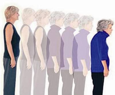
绝经后妇女骨质疏松症
相关药品
简体中文
简体中文